A couple of years ago (feels more like a whole century) when I was in high school, my eyes started giving me problems. The occasional headaches became more frequent coupled with photosensitivity, eye itching, and, eye redness. It began to affect my daily lifestyle and my studying habits. My parents were left with no other choice other than to schedule an appointment with an ophthalmologist who would then become my permanent ophthalmologist.
Fast-forward to a few days later, mum and I were in his consulting rooms, waiting for my appointment, a few minutes later it was my turn, yikes!! I remember walking into the examining room, indulging in a brief 10-minute introductory conversation, followed by explaining all my symptoms to him. Next up was the tonometry test to measure the pressure inside my eyes which revealed that my right eye pressure was elevated by two, and my left eye had decreased eye pressure by 0.5. I mean talk about opposites attracting!!!
Sometimes life is eye-ronic!! The right eye was a cause of concern and that’s when I was first introduced to the term, ‘glaucoma’. We had to get on top of the situation and it meant being prescribed steroid eye drops to regulate the eye pressure before it lead to glaucoma. I used the eye drops diligently and a couple of months later, all was back to normal. But, given my issue with elevated eye pressure, I still need to do regular eye checks with my ophthalmologist just to be in the clear. Enough about me let’s dive into the topic at hand..
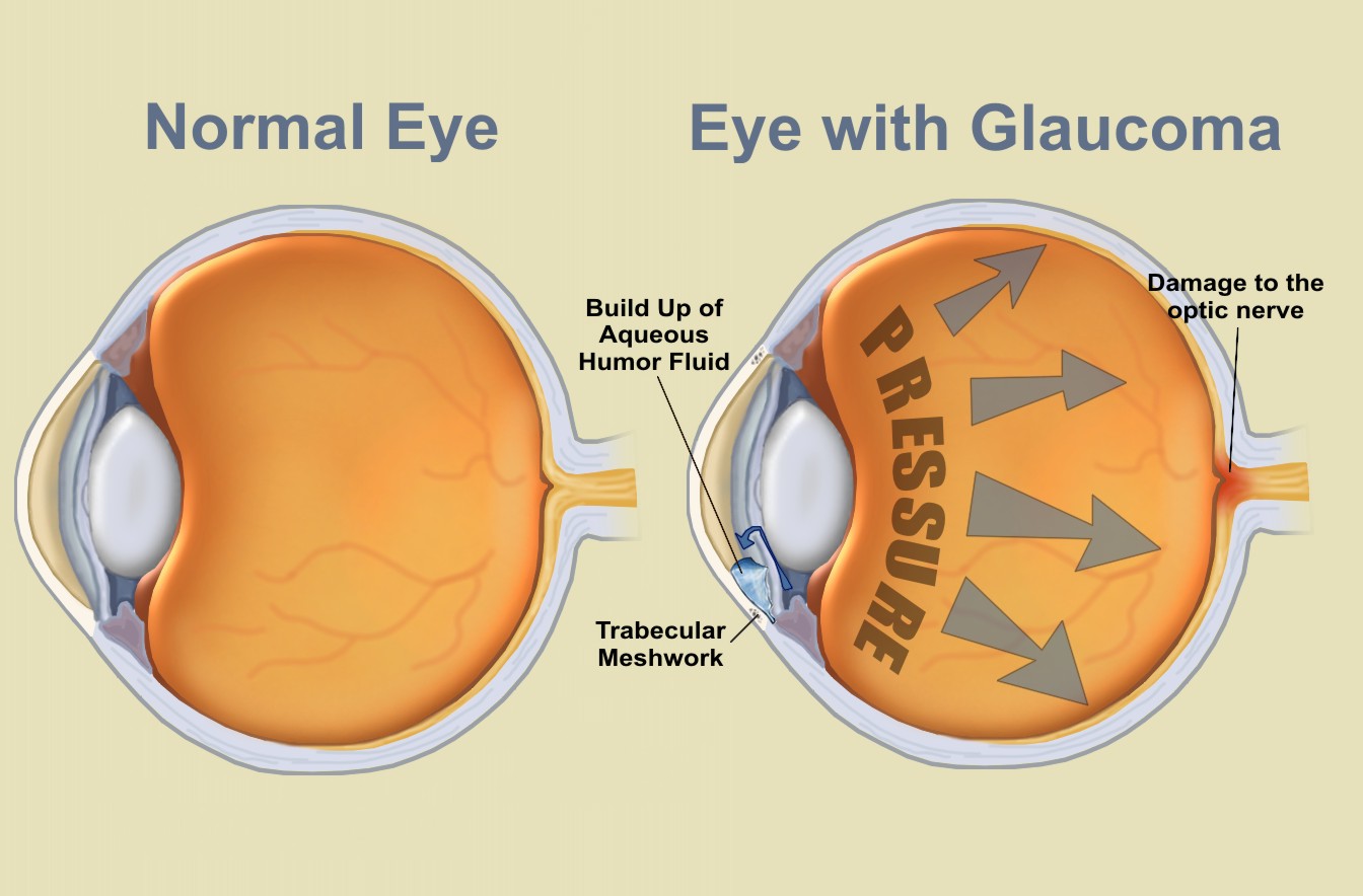
Glaucoma is defined as a group of eye conditions that damage the optic nerve, the health of which is critical for good vision. It is the most common type of optic nerve damage that results in vision loss. In most cases, fluid accumulates in the front part of the eye. This extra fluid puts pressure on the eye, gradually causing optic nerve damage. This pressure is known as intraocular pressure (IOP) or eye pressure. Glaucoma can happen at any age, but it is more common in older people.
Causes
Glaucoma tends to run in families. Scientists have discovered genes linked to high eye pressure and optic nerve damage in some people.
It is caused by optic nerve damage. Blind spots develop in your visual field as this nerve gradually deteriorates. This nerve damage is typically associated with increased eye pressure for reasons that doctors do not fully understand.
Elevated eye pressure is caused by a buildup of a fluid (aqueous humor) that flows throughout the inside of your eye. This internal fluid normally drains through a tissue called the trabecular meshwork at the angle where the iris and cornea meet.
With glaucoma, the drainage canals become clogged with microscopic deposits. Because there is nowhere for the fluid to go, it accumulates in the eye. This excess fluid puts pressure on the eye. This increased eye pressure can eventually damage the optic nerve, leading to glaucoma.
Types
- Open-angle glaucoma
The most common type of glaucoma is open-angle glaucoma. The drainage angle formed by the cornea and iris is unobstructed, but the trabecular meshwork is partially blocked. As a result, the pressure in the eye gradually rises. The optic nerve is damaged as a result of this pressure. It happens so slowly that you may lose sight before you realize there’s a problem.

2. Angle-closure glaucoma
Angle-closure glaucoma, also known as closed-angle glaucoma, is caused by the iris bulging forward, narrowing or blocking the drainage angle formed by the cornea and iris. As a result, fluid is unable to circulate through the eye, and pressure rises. Some people have narrow drainage angles, which puts them at risk of angle-closure glaucoma.
This type can develop suddenly (acute angle-closure glaucoma) or gradually (progressive angle-closure glaucoma) (chronic angle-closure glaucoma). Acute angle-closure glaucoma is a life-threatening condition.

3. Normal-tension glaucoma
Even though your eye pressure is within the normal range, your optic nerve is damaged in normal-tension glaucoma. Nobody knows for certain why this is happening. You may have a sensitive optic nerve, or you may have less blood supply to your optic nerve. This restricted blood flow could be caused by atherosclerosis (the buildup of fatty deposits (plaque) in the arteries) or other conditions that impair circulation.

4. Pigmentary glaucoma
Pigment granules from your iris accumulate in the drainage channels of your eye in pigmentary glaucoma, slowing or blocking fluid exiting your eye. Jogging, for example, can cause the pigment granules to be stirred up, depositing them on the trabecular meshwork and causing intermittent pressure elevations.

5. Congenital glaucoma
Some babies are born with drainage canals that did not form properly in the womb. Drainage blockages or an underlying medical condition could be the cause of optic nerve damage. Glaucoma symptoms in a baby can be noticed at birth or develop later during childhood. Childhood, infantile, or pediatric glaucoma are other names for this condition.

Symptoms
Signs and symptoms differ depending on the type and stage of your condition. As an example:
Open-angle glaucoma:
- Patchy blind spots in your side (peripheral) or central vision, frequently in both eyes
- Tunnel vision in the advanced stages
Acute angle-closure glaucoma:
- Severe headache
- Eye pain
- Nausea and vomiting
- Blurred vision
- Halos around lights
- Eye redness
With any type, you may experience:
- Eye pain or pressure
- Headaches
- Rainbow-colored halos around lights
- Low vision, blurred vision, narrowed vision (tunnel vision) or blind spots
- Nausea and vomiting
- Red eyes

If left untreated, glaucoma will eventually lead to blindness. Even with treatment, approximately 15% of people will go blind in at least one eye within 20 years.
Risk factors
Glaucoma can affect people of all races and genders, but the risk increases with age. African Americans and Latinos are much more likely than other races to develop glaucoma, and they tend to develop the disease at a younger age. Asian and Inuit populations are also more prone to a specific type of glaucoma, angle-closure glaucoma.
Because chronic forms of glaucoma can destroy vision before any signs or symptoms become apparent, be aware of the following risk factors:
- Having high internal eye pressure (intraocular pressure)
- Being over age 60
- Being black, Asian or Hispanic
- Having a family history of glaucoma
- Having certain medical conditions, such as diabetes, heart disease, high blood pressure and sickle cell anemia
- Having corneas that are thin in the center
- Nearsightedness known as myopia (for open-angle glaucoma)
- Farsightedness known as hyperopia (for closed-angle glaucoma)
- Previous eye injury or certain types of eye surgery
- Long-term use of corticosteroids

Diagnosis
It is possible to have glaucoma and be completely unaware of it. Regular eye exams are critical for detecting glaucoma and other vision problems. Optic health and vision loss can be assessed during an eye exam.
To check for glaucoma, your eye doctor may do one or more of these painless tests:
- Dilated eye exam to widen pupils and view the optic nerve at the back of the eyes
- Gonioscopy to examine the angle where the iris and cornea meet
- Optical coherence tomography (OCT) to look for changes in the optic nerve that may indicate glaucoma
- Ocular pressure test (tonometry) to measure eye pressure
- Pachymetry to measure corneal thickness
- Slit-lamp exam to examine the inside of the eye with a special microscope called a slit lamp
- Visual acuity test (eye charts) to check for vision loss
- Visual field test(perimetry) to check for changes in peripheral vision (your ability to see things off to the side)

Treatment
Glaucoma can’t be reversed. However, treatment and regular checkups can help slow or prevent vision loss, especially if the disease is detected early on.
It is treated by lowering eye pressure (intraocular pressure). Depending on the circumstances, options may include prescription eye drops, oral medications, laser treatment, surgery, or a combination of any of these.
a) Eye drops
Eyedrops are the first line of treatment. These can help lower eye pressure by improving how fluid drains from the eye or by reducing the amount of fluid produced by the eye. Depending on how low the eye pressure must be, more than one of the eyedrops listed below may be prescribed.
Prescription eyedrop medications include:
- Prostaglandins. These increase the outflow of the fluid in the eye (aqueous humor), thereby reducing eye pressure. Medicines in this category include latanoprost (Xalatan), travoprost (Travatan Z), tafluprost (Zioptan), bimatoprost (Lumigan) and latanoprostene bunod (Vyzulta).
Possible side effects include; mild reddening and stinging of the eyes, darkening of the iris, darkening of the pigment of the eyelashes or eyelid skin, and blurred vision. This class of drug is prescribed for once-a-day use.
- Beta blockers. These reduce the production of fluid in the eye, thereby lowering the pressure in the eye (intraocular pressure). Examples include timolol (Betimol, Istalol, Timoptic) and betaxolol (Betoptic).
Possible side effects include; difficulty breathing, slowed heart rate (bradycardia), lower blood pressure(hypotension), impotence, and fatigue. This class of drug can be prescribed for once- or twice-daily use depending on your condition.
- Alpha-adrenergic agonists. These reduce the production of aqueous humor and increase outflow of the fluid in the eye. Examples include apraclonidine (Iopidine) and brimonidine (Alphagan P, Qoliana).
Possible side effects include; an irregular heart rate, high blood pressure(hypertension), fatigue, red, itchy or swollen eyes, and dry mouth. This class of drug is usually prescribed for twice-daily use but sometimes can be prescribed for use three times a day.
- Carbonic anhydrase inhibitors. These medicines reduce the production of fluid in the eye. Examples include dorzolamide (Trusopt) and brinzolamide (Azopt). Possible side effects include; a metallic taste, frequent urination, and tingling in the fingers and toes. This class of drug is usually prescribed for twice-daily use but sometimes can be prescribed for use three times a day.
- Rho kinase inhibitor. This medicine lowers eye pressure by suppressing the rho kinase enzymes responsible for fluid increase. It is available as netarsudil (Rhopressa) and is prescribed for once-a-day use. Possible side effects include; eye redness, eye discomfort and deposits forming on the cornea.
- Miotic or cholinergic agents. These increase the outflow of fluid from the eye. An example is pilocarpine (Isopto Carpine). Side effects include; headache, eye ache, smaller pupils, possible blurred or dim vision, and nearsightedness. This class of medicine is usually prescribed to be used up to four times a day. Because of potential side effects and the need for frequent daily use, these medications are not prescribed very often anymore.
Since some of the eyedrop medication is absorbed into the bloodstream, some side effects unrelated to the eyes may occur. Close your eyes for one to two minutes after putting the drops in to reduce absorption. You can also press lightly at the corner of your eyes near your nose for one or two minutes to close the tear duct. Remove any unused drops from your eyelid by gently wiping them off.
If you have been prescribed multiple eyedrops or need to use artificial tears, space them out so that you wait at least five minutes between types of drops.
b) Oral medications
If eyedrops alone are ineffective in lowering your eye pressure, your doctor may prescribe an oral medication, usually a carbonic anhydrase inhibitor. Frequent urination, tingling in the fingers and toes, depression, stomach upset, and kidney stones are all possible side effects.
c) Surgery and other therapies
Other treatment options, in addition to eye drops and oral medications, include laser therapy and various surgical procedures. The following techniques are intended to improve fluid drainage within the eye and thus lower pressure:
- Laser therapy. This is performed in your doctor’s office. Laser trabeculoplasty is an option if you have open-angle glaucoma. A small laser beam is used to open clogged channels in the trabecular meshwork. It may take a few weeks before the full effect of this procedure becomes apparent.
- Filtering surgery. With a surgical procedure called a trabeculectomy, your surgeon creates an opening in the white of the eye (sclera) and removes part of the trabecular meshwork.
- Drainage tubes. In this procedure, your eye surgeon inserts a small tube shunt in your eye to drain away excess fluid to lower your eye pressure.
- Minimally invasive glaucoma surgery (MIGS). Your doctor may suggest a MIGS procedure to lower your eye pressure. These procedures generally require less immediate postoperative care and have less risk than trabeculectomy or installing a drainage device. They are often combined with cataract surgery. There are a number of MIGS techniques available, and your doctor will discuss which procedure may be right for you.
Post-procedure follow-up exams are an important part of the journey. If your eye pressure begins to rise or other changes occur in your eye, you may eventually need to undergo additional procedures.
Treating acute angle-closure glaucoma
Acute angle-closure glaucoma is a medical emergency. If you are diagnosed with this condition, you will require immediate treatment to alleviate the pressure in your eye. In most cases, this will necessitate the use of medication as well as laser or other surgical procedures.
A laser peripheral iridotomy is a procedure in which the doctor uses a laser to create a small opening in your iris. This allows fluid (aqueous humor) to circulate through it, relieving eye pressure.
d) Home remedies and lifestyle modification
The following suggestions may assist you in controlling high eye pressure or promoting eye health:
- Eat a healthy diet. Eating a healthy diet can help you maintain your health, but it won’t prevent glaucoma from worsening. Several vitamins and nutrients are important to eye health, including zinc, copper, selenium, and antioxidant vitamins C, E, and A.
- Exercise safely. Regular exercise may reduce eye pressure in open-angle glaucoma. Talk to your doctor about an appropriate exercise program.
- Limit your caffeine. Drinking beverages with large amounts of caffeine may increase your eye pressure.
- Sip fluids frequently. Drink only moderate amounts of fluids at any given time during the course of a day. Drinking a quart or more of any liquid within a short time may temporarily increase eye pressure.
- Sleep with your head elevated. Using a wedge pillow that keeps your head slightly raised, about 20 degrees, has been shown to reduce intraocular pressure while you sleep.
- Take prescribed medicine. Using your eyedrops or other medications as prescribed can help you get the best possible result from your treatment. Be sure to use the drops exactly as prescribed. Otherwise, your optic nerve damage could worsen.
- Relaxation techniques. Stress may trigger an attack of acute angle-closure glaucoma. If you’re at risk of this condition, find healthy ways to cope with stress. Meditation and other techniques may help.

Prevention
Detection of glaucoma in its early stages is important in preventing vision loss or slowing its progress. Below are effective ways of staying on top of the glaucoma issue:
- Get regular dilated eye examinations. Regular comprehensive eye exams can help detect glaucoma in its early stages, before significant damage occurs. As a general rule, the American Academy of Ophthalmology recommends having a comprehensive eye exam every;
- five to 10 years if you’re under 40 years old;
- two to four years if you’re 40 to 54 years old;
- one to three years if you’re 55 to 64 years old;
- one to two years if you’re older than 65.
- six to twelve months if you’re at risk of glaucoma.
- Know your family’s eye health history. Glaucoma tends to run in families. If you’re at increased risk, you may need more frequent screening.
- Exercise safely. Regular, moderate exercise may help prevent glaucoma by reducing eye pressure. Talk with your doctor about an appropriate exercise program.
- Take prescribed eyedrops regularly. Glaucoma eyedrops can significantly reduce the risk that high eye pressure will progress to glaucoma. To be effective, eyedrops prescribed by your ophthalmologist need to be used regularly even if you have no symptoms.
- Wear eye protection. Serious eye injuries can lead to glaucoma. Wear eye protection when using power tools or playing high-speed racket sports in enclosed courts.

Prognosis
Blindness is a rare complication of glaucoma that can be prevented if detected early. On the other hand, glaucoma is a chronic and progressive condition that frequently results in some degree of vision loss over time. The earlier you detect it and begin treatment, the better your chances of preserving your vision. Treatments can slow the progression of the disease and prevent vision loss. Regular eye exams are essential if you are at high risk for glaucoma.

To summarize, while there is no cure for glaucoma, treatments can keep eye pressure under control and vision loss at bay. Eye exams can detect the disease early and help you keep your sight. If you have a high risk of glaucoma, ask your eye doctor (ophthalmologist) how frequently you should have screenings. If you have glaucoma, you must use daily eye drops as directed. Inquire with your ophthalmologist about laser treatments and surgical options. You can prevent glaucoma from worsening and causing irreversible vision loss or blindness if you take proper care of it.
That’s all I’ve got for today. Who knew our eyes had more issues than Vogue!!


Leave a Reply