ENDOMETRIOSIS

Home Health ENDOMETRIOSIS
ENDOMETRIOSIS

The uterus also called the womb is a hollow, pear-shaped muscular organ of the female reproductive system found in the pelvic area between the bladder and the rectum.  It is usually the size of a fist. The uterus is connected to the fallopian tubes (responsible for transferring eggs from your ovaries into the uterus). The cervix is the lower part of the uterus and connects to the vagina. The corpus or fundus is the wider, upper part of the uterus.

Women's reproductive anatomy - A simple guide - Learn more here

The uterus consists of 3 layers:

  • Endometrium: this is the inner lining and it is shed during your period.
  • Myometrium: this is the thick middle muscle layer of the corpus or fundus. This expands during pregnancy to hold the growing baby. During labor, it contracts to push the baby out.
  • Serosa: this is the smooth outer layer. It covers the uterus and makes mobility of the uterus easy.

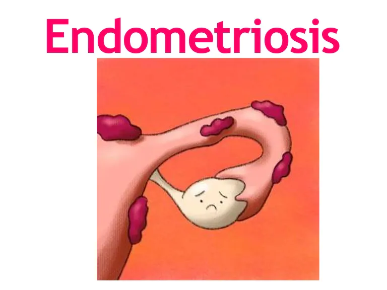
Endometriosis happens when cells of the endometrium (endometrial tissue) grow outside the uterus. Common locations of endometriotic implants are:

  1. Peritoneum
  2. Pelvic organs: ovaries (most common & usually bilateral), fallopian tubes, bladder, cervix, rectouterine pouch.
  3. Extra-pelvic organs: diaphragm, lungs, bowel, laparotomy scars.
What is Endometriosis?

The cause of endometriosis is not yet fully understood. Risk factors include:

  • a family history of the condition
  • retrograde menstruation (backflow of menstrual fluid containing endometrial cells)
  • coelomic metaplasia
  • iatrogenic implantation (postsurgical scars through episiotomy or laparotomy as prime locations)
  • hematogenic (blood related) dissemination of endometrial cells
Risk factors in association with endometriosis. | Download Scientific  Diagram

Approximately one-third of women with endometriosis are asymptomatic. When symptoms do occur, they tend to reflect the area of involvement:

  • dysmenorrhea
  • heavy, irregular bleeding (pre-or postmenstrual bleeding)
  • pelvic pain
  • dyspareunia (pain during or after sex)
  • pain with bowel movement (dyschezia)
  • infertility (50% of patients)
Endometriosis Symptoms - EndoStats - Endometriosis Awareness

We have covered the definition, commonly affected sites, risk factors, and symptoms, now let us talk about how to diagnose it.

Diagnosis;

  1. Patient history
  2. Physical examination. This will reveal rectovaginal tenderness and adnexal masses.
  3. The primary diagnostic modality for endometriosis is Laparoscopy. This is a surgical procedure where a camera is used to look inside the abdominal cavity and may show endometriotic implants and adhesions.
  4. Transvaginal ultrasound. This shows evidence of ovarian cysts (chocolate cysts)
  5. MRI (magnetic resonance imaging)

NB: NORMALLY THE SEVERITY OF THE FINDINGS DOES NOT CORRELATE WITH THE SEVERITY OF SYMPTOMS.

How Endometriosis Is Diagnosed

In 1997, the American Society of Reproductive Medicine revised the surgical classification of endometriosis into 4 stages;

Stage 1 (minimal): findings restricted to only superficial lesions and a few filmy adhesions.

Stage 2 (mild): in addition, some deep lesions are present in the cul-de-sac.

Stage 3 (moderate): as above, plus the presence of endometriomas on the ovary and more adhesions.

Stage 4 (severe): as above, plus large endometriomas, extensive adhesions.

Endometriosis and Adenomyosis | Obgyn Key

There is no cure for endometriosis. But two types of interventions are possible:

  1. Treatment of pain
  2. Treatment of endometriosis-associated infertility.

The main goal is to provide pain relief, restrict progression of the process and where needed, restore or preserve fertility.

Since endometriosis is dependent on the cyclic production of menstrual cycle hormones, this can be used as the basis for medical therapy.

For mild-moderate pelvic pain not associated with complications;

  • Nonsteroidal-Anti-Inflammatory drugs (NSAIDs) and continuous hormonal contraceptives (Estrogen-Progestin or Progestogen-only)
  • Progestational agents (Norethindrone)
  • Synthetic androgens (Danazol)

For severe symptoms;

  • GnRH agonists (Buserelin, Goserelin)
  • Estrogen-Progestin oral contraceptive pills.

Surgical care can be classified as follows:

A. Conservative- when the reproductive potential is retained.

  • Drainage and laparoscopic cystectomy
  • Ablation
  • Presacral neurectomy
  • Laparoscopic uterine nerve ablation (LUNA)

B. Semiconservative- when the reproductive ability is eliminated but the ovarian function is retained. This is mainly indicated for:

  • women who have finished giving birth
  • are too young for surgical menopause
  • who have severe symptoms and are debilitated by them.

Hysterectomy and cytoreduction of pelvic endometriosis are the surgeries of choice for this group.

C. Radical- when the uterus and ovaries are removed.

Radical surgery involves total hysterectomy with bilateral oophorectomy (TAH-BSO) and cytoreduction of visible endometriosis.

Treatment — Nuffield Department of Women's & Reproductive Health

Adhesiolysis is performed to restore mobility and normal intrapelvic organ relationships.

That is all for now. Catch y’all later (‘-‘)

Leave a Reply

Your email address will not be published.